lv enlargement radiopaedia | elevated left ventricular volume x ray lv enlargement radiopaedia The parasternal long axis and apical four-chamber views on transthoracic echocardiography are often the primary views used to gain both a qualitative and quantitative . My pick for this list was the first one I put down for this list. There is only one vintage Omega that is higher on my list, and that is a Speedmaster Professional 145.022-76 from 1977, my year of birth. But as I mentioned, we have the Speedy Tuesday articles for the . See more
0 · normal left ventricular enlargement
1 · left ventricular enlargement ultrasound
2 · left ventricular enlargement radiology
3 · left ventricular enlargement pictures
4 · left ventricular enlargement morphology
5 · left ventricular enlargement ct
6 · elevated left ventricular volume x ray
7 · echocardiogram left ventricular enlargement
The Rolex Military Submariner. In 1972 the Rolex Submariner replaced the Omega Seamaster as the watch of choice for its the UK military by the Ministry of Defence .
The parasternal long axis and apical four-chamber views on transthoracic echocardiography are often the primary views used to gain both a qualitative and quantitative .Left atrial enlargement (LAE) may result from many conditions, either congenital .
Left ventricular enlargement can be reliably identified on nongated contrast-enhanced multidetector CT when the maximum luminal diameter of the LV is greater than 5.6 cm. . Left ventricular hypertrophy (LVH) is generally considered to be an adaptive response that allows for normal ejection fraction despite abnormal pressure and/or volume . LV enlargement (LVE) is associated with a wide variety of cardiovascular processes, including cardiomyopathy, myocardial infarction, valvular heart disease, PH, and .
LV mass assessed by echocardiography and CMR, cardiovascular outcomes, and medical practice. JACC Cardiovasc Imaging 2012 ;5(8):837–848. Medline Google Scholar Left ventricular hypertrophy was strongly associated with hard coronary heart disease events (death and myocardial infarction) over 15 years of follow-up in a contemporary . Radiological sign of left ventricular enlargement based on the distance between the inferior vena cava (IVC) and left ventricle (LV). Left ventricular enlargement is suggested on lateral chest radiograph if the . Left atrial enlargement (LAE) may result from many conditions, either congenital or acquired. It has some characteristic findings on a frontal chest radiograph. CT or MRI may .
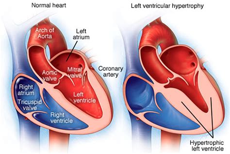
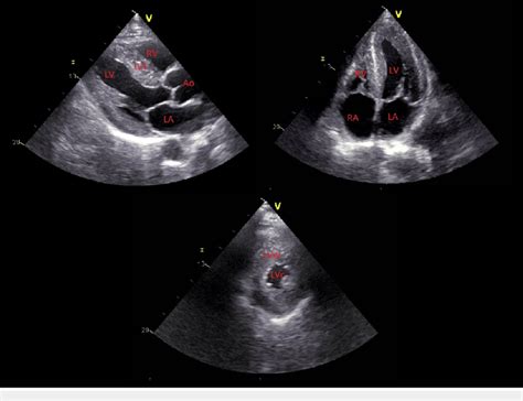
Normal LV myocardial thickness is <11 mm. 5 LVH is considered mild if it measures 11-13 mm, moderate if it measures 14-15 mm, and severe if it measures >15 mm. LVH can . The parasternal long axis and apical four-chamber views on transthoracic echocardiography are often the primary views used to gain both a qualitative and quantitative appreciation of left ventricular enlargement. Features include 4: increased left ventricular internal end-diastolic diameter (LVIDd)
Left ventricular hypertrophy is a condition where the muscle wall of the heart's left pumping chamber thickens.Left ventricular enlargement can be reliably identified on nongated contrast-enhanced multidetector CT when the maximum luminal diameter of the LV is greater than 5.6 cm. Nongated contrast-enhanced CT scan can be used to recognize LVE. Left ventricular hypertrophy (LVH) is generally considered to be an adaptive response that allows for normal ejection fraction despite abnormal pressure and/or volume load. 1 However, this adaptation is associated with increased cardiac morbidity and mortality, including acute myocardial infarction, heart failure, arrhythmia, and stroke. 2–4 .
LV enlargement (LVE) is associated with a wide variety of cardiovascular processes, including cardiomyopathy, myocardial infarction, valvular heart disease, PH, and congestive heart failure (25–28). LV mass assessed by echocardiography and CMR, cardiovascular outcomes, and medical practice. JACC Cardiovasc Imaging 2012 ;5(8):837–848. Medline Google Scholar

Left ventricular hypertrophy was strongly associated with hard coronary heart disease events (death and myocardial infarction) over 15 years of follow-up in a contemporary ethnically diverse cohort, after adjustment for traditional risk factors and coronary artery calcium. Radiological sign of left ventricular enlargement based on the distance between the inferior vena cava (IVC) and left ventricle (LV). Left ventricular enlargement is suggested on lateral chest radiograph if the distance (A) between the LV border border and the posterior border of IVC is more than 1.8 cm at a level 2 cm above the intersection of . Left atrial enlargement (LAE) may result from many conditions, either congenital or acquired. It has some characteristic findings on a frontal chest radiograph. CT or MRI may also be used for diagnosis.
normal left ventricular enlargement
Normal LV myocardial thickness is <11 mm. 5 LVH is considered mild if it measures 11-13 mm, moderate if it measures 14-15 mm, and severe if it measures >15 mm. LVH can also be defined and quantified in terms of absolute and BSA-indexed LV mass values, with values over the 95th percentile considered abnormal (91 g/m 2 in males and 77 g/m 2 in . The parasternal long axis and apical four-chamber views on transthoracic echocardiography are often the primary views used to gain both a qualitative and quantitative appreciation of left ventricular enlargement. Features include 4: increased left ventricular internal end-diastolic diameter (LVIDd)
chanel shoes winter 2022
Left ventricular hypertrophy is a condition where the muscle wall of the heart's left pumping chamber thickens.Left ventricular enlargement can be reliably identified on nongated contrast-enhanced multidetector CT when the maximum luminal diameter of the LV is greater than 5.6 cm. Nongated contrast-enhanced CT scan can be used to recognize LVE.
Left ventricular hypertrophy (LVH) is generally considered to be an adaptive response that allows for normal ejection fraction despite abnormal pressure and/or volume load. 1 However, this adaptation is associated with increased cardiac morbidity and mortality, including acute myocardial infarction, heart failure, arrhythmia, and stroke. 2–4 . LV enlargement (LVE) is associated with a wide variety of cardiovascular processes, including cardiomyopathy, myocardial infarction, valvular heart disease, PH, and congestive heart failure (25–28). LV mass assessed by echocardiography and CMR, cardiovascular outcomes, and medical practice. JACC Cardiovasc Imaging 2012 ;5(8):837–848. Medline Google Scholar Left ventricular hypertrophy was strongly associated with hard coronary heart disease events (death and myocardial infarction) over 15 years of follow-up in a contemporary ethnically diverse cohort, after adjustment for traditional risk factors and coronary artery calcium.
Radiological sign of left ventricular enlargement based on the distance between the inferior vena cava (IVC) and left ventricle (LV). Left ventricular enlargement is suggested on lateral chest radiograph if the distance (A) between the LV border border and the posterior border of IVC is more than 1.8 cm at a level 2 cm above the intersection of . Left atrial enlargement (LAE) may result from many conditions, either congenital or acquired. It has some characteristic findings on a frontal chest radiograph. CT or MRI may also be used for diagnosis.
left ventricular enlargement ultrasound

left ventricular enlargement radiology
1982 Rolex Day Date. Save Search. Malachite Rolex. Rolex 19018. Rolex Gmt Master 16750. Rolex 15000. Rolex 18078. Rolex With Lapis. Rolex Wood Dial. Cotton Bowl Classic. More Popular Searches. 1982 Rolex President Day-Date Wristwatch Made in Yellow Gold, Ref. 18038. Free Shipping.
lv enlargement radiopaedia|elevated left ventricular volume x ray


